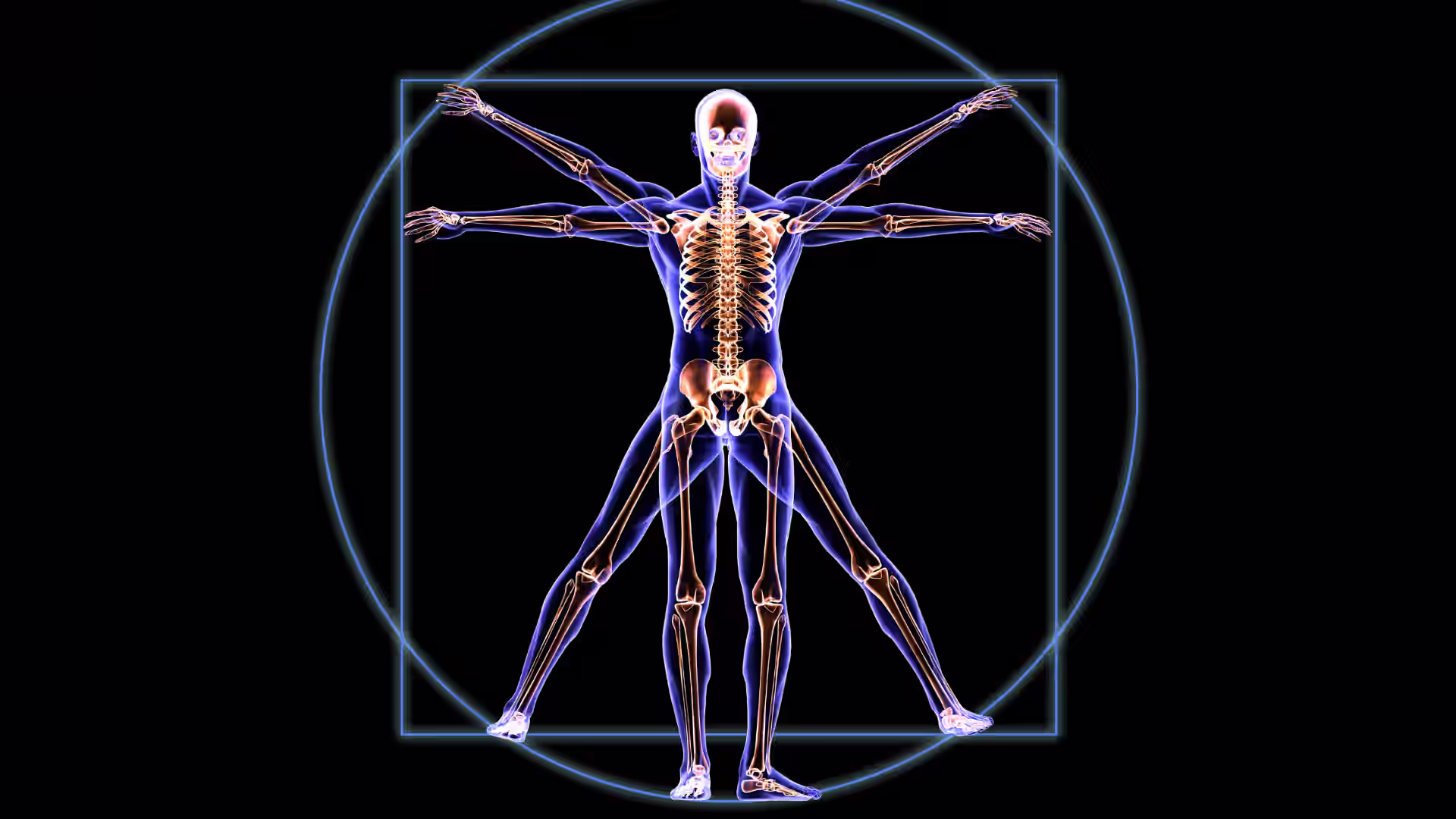


Dermatomes are specific areas of skin that receive sensory input—such as touch, pain, and temperature—from a single spinal nerve. These mapped regions help healthcare professionals assess nerve function and identify potential injuries or conditions affecting the nervous system.
Understanding dermatomes is essential for diagnosing nerve-related issues, including spinal cord disorders and conditions like shingles. Dermatome mapping serves as a valuable tool in pinpointing affected nerves and guiding treatment decisions.
This guide provides a clear overview of dermatomes, their medical significance, and how they are used in clinical practice.
[signup]
What Are Dermatomes?
Dermatomes are like a sensory map on your skin. Each dermatome is connected to a specific spinal nerve, which sends signals from that area of skin to your brain.
Each spinal nerve corresponds to a specific skin region, transmitting sensory signals from that area to the brain.
Connecting Skin to Nerves
Every time you touch something, feel the temperature or get a pain signal, it travels from your skin through these nerves to your brain. Dermatomes help doctors understand which nerve is sending the signals. This connection is vital for diagnosing issues like nerve damage or spinal injuries.
Physiology of Touch: How Dermatomes Translate Sensation
Each spinal nerve sends signals out from the brain to the body and skin and also sends signals from the skin to the brain. Each spinal nerve primarily transmits sensations from its corresponding dermatome, though some overlap occurs.
Different types of sensors (nerve endings) transmit different signals:
Touch Receptors
Your skin has special sensors that help you feel different sensations, such as touch, pressure, and vibrations. These sensations travel through nerves that follow specific dermatomes.
Meissner corpuscles sense light touch, Pacinian corpuscles detect deep pressure and vibration, and Ruffini endings feel skin stretching. Merkel cells are specialized slow-adapting touch receptors in the skin that detect fine details, textures, and sustained pressure; they help us feel shapes and edges.
Pain and Temperature Sensors
Tiny nerve endings in the skin help you feel pain, heat, and cold. They warn your body about things that could hurt you, like touching something too hot or sharp.
These signals are also transmitted along nerves belonging to specific dermatomes.
Messages to the Brain
From each dermatome, these sensors send signals through nerves to your brain, which helps you understand what you're feeling and react quickly, like pulling your hand away from something hot.
History and Discovery of Dermatomes
The history of dermatomes shows how our understanding of these skin nerve maps has changed over time:
First Maps (Late 1800’s–Early 1900’s)
Scientists began studying dermatomes in the late 1800s. During that time, scientists like Herringham, Head, and Sherrington were among the first to map dermatomes.
Using cadaver dissections, clinical observations (such as shingles rashes), and experiments on monkeys, they figured out which nerves controlled different areas of skin.
A scientist, neurosurgeon, and prolific writer named Otfrid Foerster later expanded this work by cutting spinal nerve roots (rhizotomy) and mapping which areas lost sensation. He published his work in the paper "The Dermatomes in Man."
More Research (1948)
J. Keegan and F. Garrett added to this knowledge by studying how spinal nerves send signals to different parts of the body. They published their findings in "The Segmental Distribution of the Cutaneous Nerves in the Limbs of Man."
Updated Understanding (2008)
Scientists M. Lee, R. McPhee, and M. Stringer questioned the older dermatome maps and suggested a more accurate way to map them using new research. They published their findings in "An Evidence-Based Approach to Human Dermatomes."
This history shows that while classic dermatome maps are helpful, scientists are still learning more about how nerves in the skin connect to the spinal cord.

Dermatome Map Explained
To understand a visual dermatome map, imagine the body divided into skin regions, each controlled by a specific spinal nerve. These maps show how sensory signals travel from the skin to the spinal cord and brain.
Each dermatome is labeled with a letter and number, such as C6 (cervical), T4 (thoracic), L4 (lumbar), or S1 (sacral), which correspond to the spinal nerve that serves that area on the skin.
Key Points for Reading a Dermatome Map:
- Cervical (C1-C8): covers the head, neck, shoulders, arms, and hands.
- Thoracic (T1-T12): covers the chest and mid-back.
- Lumbar (L1-L5): covers the lower back, thighs, knees, and parts of the feet.
- Sacral (S1-S5): covers the back of the legs, buttocks, and pelvic region.
The map helps diagnose nerve issues by matching numbness, pain, or tingling areas to the affected spinal nerve. However, because dermatomes overlap, different maps may show slight variations.
Clinical Uses of Dermatome Maps
Dermatome maps assist in assessing nerve function, but they should be used alongside other diagnostic tools like imaging and nerve conduction studies.
Diagnosing Nerve Damage
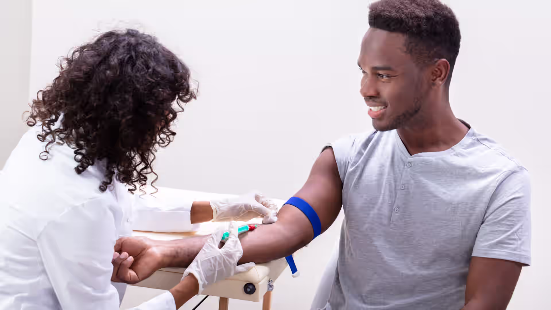
Patients with nerve damage might feel pain, numbness, or tingling in specific areas. Based on symptom location, dermatome maps can help doctors assess potential spinal nerve involvement, though further testing is typically needed.
Tracking Conditions Like Shingles
Shingles, caused by the reactivation of the chickenpox virus, often follows a dermatome. Because the virus remains in nerves after infection, the rash and pain usually appear in the dermatomes associated with the nerve containing a reactivated virus.
Diagnosing Neurological Conditions: How Doctors Test Dermatome Functionality
Doctors use sensory tests to assess nerve function in different dermatomes, such as:
- Pinprick Test: this test involves using a small needle or sharp object to see if the patient can feel sharp pain along different dermatomes.
- Light Touch Test: gently touching the skin to check for sensations.
- Temperature Test: applying warm or cold objects to assess temperature sensitivity along dermatomes.
Symptoms Indicating Dermatome Issues
If a patient feels numbness, tingling, burning, or unusual pain in a specific area, it might indicate a nerve problem with the corresponding dermatome. These symptoms help doctors narrow down the location of nerve damage or disease.
Targeted Treatments Using Dermatome Knowledge
With knowledge of dermatomes, doctors can confirm diagnoses and consider targeted treatments like nerve blocks or steroid injections, though effectiveness varies by individual. This workup ensures that the treatment reaches the exact area causing the pain.
Treatment effectiveness varies; possible treatments should always be discussed with your healthcare provider.
Using Dermatomes in Regional Anesthesia
Dermatome maps assist anesthesiologists in planning regional anesthesia, along with other patient-specific factors.
Epidural and Spinal Anesthesia Mapping
Anesthesiologists use dermatome maps to determine where to apply anesthesia for surgeries.
Nerve Blocks and Dermatomes
Peripheral nerve blocks are a type of pain relief where doctors inject numbing medicine near specific nerves to block pain signals along certain dermatomes of the body.
Variations in Dermatome Maps
While incredibly helpful, dermatomes are not entirely universal from person to person. They can vary between individuals because spinal nerves overlap. Think of each spinal nerve as a tree trunk that branches into a network of roots. As these roots spread, they become finer and start to intertwine. Similarly, spinal nerves extend outward and overlap as they travel toward the extremities, meaning some areas of the body may receive more input from one nerve than another.
Challenges in Clinical Practice
While dermatomes provide excellent clinical insight, further testing is often required to identify the nerve root involved accurately.
Misdiagnosis Due to Overlapping Dermatomes
Because some dermatomes overlap, doctors might sometimes misinterpret symptoms or sensory test results, leading to incorrect diagnoses. It’s essential to use additional testing methods to confirm findings.
Improving Accuracy Through Modern Techniques
Advances in technology, such as high-resolution neuroimaging and nerve conduction studies, help doctors better understand nerve function and improve diagnostic accuracy.
Interactive Learning & Practical Applications
If you're studying dermatomes, the following activities can help you better understand how doctors use them practically.
Self-testing is for educational purposes only and is not diagnostic.
Hands-On Dermatome Testing
The following quick tests can help you better understand the dermatomes.
Self-Test Section
Try This:Ask a friend to help you test your dermatomes. Close your eyes and have them gently poke different areas of your skin. See if you can identify which dermatome each poke likely belongs to. This hands-on activity can help you understand how dermatomes work.
Quick Clinical Cases for Self-Assessment
Case 1: A patient feels numbness in their thumb. Which dermatome is likely affected?
Answer: The C6 dermatome.
Case 2: A patient has a burning sensation on the inner thigh and knee. Which dermatome is involved?
Answer: The L3 dermatome.
[signup]
Key Takeaways
- Dermatomes Help Map Sensory Signals: each dermatome corresponds to a specific spinal nerve, helping doctors assess nerve involvement in conditions like shingles, nerve damage, and spinal disorders.
- Clinical Importance of Dermatome Maps: doctors use dermatome maps to assess nerve function, guide regional anesthesia, and administer targeted treatments such as nerve blocks and steroid injections for precise pain relief.
- Dermatome Variability and Diagnostic Challenges: while dermatome maps provide valuable clinical insights, individual variations, and overlapping spinal nerve coverage can sometimes lead to misdiagnosis, requiring additional testing like nerve conduction studies for accuracy.